Neurological Assessment
(Starkey, 2010)
- If there is a suspected neurological injury, a neurological tests of the dermatomes and mytomes should be conducted. Neurological tests can be used to identify the following pathologies: nerve root impingement, peripheral nerve damage or entrapment, central nervous system trauma, or disease.
- Symptoms of a possible neurological pathology can include: numbness, tingling, paresthesia, muscular weakness, and pain.
- Neurological signs must be determined so that proper treatment and management techniques by the athletic trainer can be performed.
- To assess the integrity of the nervous system, evaluation of the dermatomes and myotomes must occur.
- To evaluate the dermatomes, have the patient differentiate between light touch discrimination: sharp and dull. Compare bilaterally. The sensations should be administered equally on both sides and the patient's eyes should be closed. This can help identify exactly where the problem begins and ends.
- To evaluate the myotomes, manual muscle testing should be performed. If weakness is shown in a neurological motor test, identify another muscle that shares the innervation and perform MMT. If only one muslce is weak, pathology to the muscle or peripheral nerve should be suspected. If both muscles are weak the the nerve root or peripheral nerve supplying the muscles is implicated.
- To evaluate the reflexes of the lower body use a reflex testing tool such as a reflex hammer. Have the patient sit hook seated and relaxed off the edge of a table. Tap the reflex hammer over the desired area and assess the bodies neurological reaction.
(Starkey, 2010)
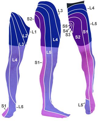
Lower Body
Dermatomes Myotomes Reflexes
L1: Groin L1: Hip flexion L1: No relfex
L2: Thigh L2: Hip flexion L2: Patellar tendon, Femoral n.
L3: Superior patella L3: Knee extension L3: Patellar tendon, Femoral n.
L4: Medial tibia/medial arch L4: Dorsiflexion and inversion L4: Patellar tendon, Femoral n.
L5: Dorsal and lateral foot L5: Great toe extension L5: Achilles Tendon, Tibial n.
S1: Plantar aspect of the foot S1: Plantarflexion S1: Achilles Tendon, Tibial n.
S2: Posterior thigh S2: Knee flexion S2: Bicep Femoris tendon,
Tibial n.
(Starkey, 2010)
http://www.google.com/imgres?q=dermatomes+lower+body&hl=en&tbo=d&biw
